Vet-written feline eye guide
Quick answer: A cat’s third eyelid may briefly appear while the cat is waking, deeply relaxed, or recovering from sedation. If it stays visible while the cat is fully awake, covers a noticeable part of one or both eyes, or appears with squinting, redness, discharge, unequal pupils, weakness, vomiting, diarrhea, or poor appetite, arrange a veterinary examination. A painful, cloudy, injured, enlarged, or suddenly abnormal eye deserves same-day care. Breathing trouble, collapse, major trauma, or a blue-gray gum color is an emergency.
Seeing a pale sheet slide across the inner corner of your cat’s eye can be unsettling. Owners often describe it as a film, extra eyelid, white skin, or pink membrane. It is the third eyelid, also called the nictitating membrane. Every cat has one in each eye. It helps protect the surface of the eye and contributes to tear distribution, but in a healthy, alert cat it is usually tucked away and barely noticeable.
The important question is not simply, “Why is my cat’s third eyelid showing?” It is how long it is showing, whether one or both eyes are affected, and what the rest of the cat is doing. A sliver seen for a few seconds after sleep is very different from a membrane that remains raised all afternoon while the cat hides and refuses food. This guide explains those patterns, the possible causes, safe observations you can make at home, and the warning signs that should move you from monitoring to veterinary care.
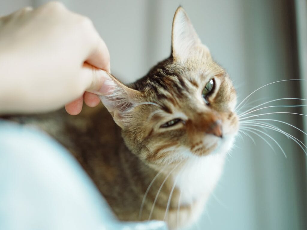
Cat third eyelid showing: What is a cat’s third eyelid?
The third eyelid is a thin, mobile fold of tissue at the inner corner of each eye. It can sweep diagonally across the eye’s surface. Unlike the upper and lower lids, you do not usually see it move during an ordinary blink. Its position is influenced by the eye’s placement in the socket, surrounding tissues, nervous-system tone, and the cat’s general physical condition.
The membrane is not the same thing as a cataract. A cataract is an opacity inside the lens, seen through the pupil. The third eyelid lies over the front of the eye and advances from the inner corner. It is also not automatically “cherry eye.” Cherry eye refers to prolapse of the tear-producing gland associated with the third eyelid, creating a rounded pink or red mass. That condition is far more common in dogs than in cats.
A tiny edge of the membrane may be visible when a cat is asleep, just waking, extremely relaxed, sedated, or anesthetized. It should normally retreat once the cat is awake and alert. Persistent elevation is a sign rather than a diagnosis. It tells us that something has changed in the eye, orbit, nervous system, hydration, or overall health.
Cat third eyelid showing: Is a visible third eyelid ever normal?
Yes, for a short time. A sleepy cat may roll the eyes slightly backward, allowing the third eyelids to rise. You might notice this when the cat wakes in your lap or opens its eyes during a nap. The same can happen after sedation or general anesthesia because normal muscle tone is temporarily reduced. In these situations the cat should otherwise look comfortable, and the membranes should gradually disappear as alertness returns.
Occasionally, owners see only a narrow pale crescent at the inner corners. If it is symmetrical, fleeting, and absent when the cat is active, it may simply be the normal anatomy becoming visible. Take a clear photo and compare it later rather than repeatedly touching the eyelids.
It is less likely to be normal when the membrane stays up while the cat is walking, eating, and looking around; covers more of the eye over time; affects one eye much more than the other; or occurs with any other sign of discomfort or illness. Kittens, seniors, and cats with chronic disease deserve a lower threshold for a veterinary call because their condition can change quickly.
Cat third eyelid showing: When a cat’s third eyelid showing is an emergency
Seek urgent or emergency veterinary care if you see any of the following:
- Severe squinting, the eye held shut, or obvious eye pain
- A cloudy, blue, bulging, bleeding, cut, punctured, or displaced eye
- Sudden vision loss, bumping into objects, panic, or disorientation
- Unequal pupils with a new head tilt, weakness, circling, or collapse
- Known trauma, a cat fight, chemical splash, or foreign object near the eye
- Repeated vomiting or diarrhea with marked lethargy or dehydration
- Open-mouth breathing, severe breathing effort, or gray-blue gums
Eye emergencies are time-sensitive because the cornea and deeper structures can deteriorate rapidly. If a chemical has splashed into the eye, contact a veterinarian or animal poison service immediately and follow their flushing instructions. Do not delay care to take multiple photographs or try leftover medication. For broader crisis preparation, keep our emergency first-aid guide for dogs and cats bookmarked.
Cat third eyelid showing: Common reasons a cat’s third eyelid is showing
Cat third eyelid showing: 1. Sleep, sedation, or deep relaxation
This is the reassuring pattern: both membranes appear briefly, the eyes are not red or painful, and they retract as the cat wakes. After anesthesia, the clinic should explain what recovery signs are expected. Call if the membrane remains prominent beyond the advised recovery period or if the cat is unusually dull, cold, vomiting, or breathing abnormally.
2. Eye pain or surface irritation
A painful eye may retract slightly into the socket, allowing the third eyelid to move forward. Corneal scratches and ulcers, conjunctivitis, a trapped seed or hair, eyelid problems, and trauma can all produce this response. Typical companions are squinting, excessive tearing, pawing at the face, light sensitivity, discharge, or redness.
A corneal ulcer cannot be reliably diagnosed by looking at a photo. Veterinarians commonly apply fluorescein dye, which highlights damage to the corneal surface. They may also measure tear production and eye pressure. These tests help separate problems that can look similar but require very different medication.
3. Conjunctivitis or respiratory infection
Inflammation of the conjunctiva—the pink tissue around the eye—can make the third eyelid swollen and visible. In cats, conjunctivitis may be associated with infectious agents, irritants, allergies, or other eye disease. Sneezing, nasal discharge, mouth ulcers, fever, or reduced appetite may point to a broader respiratory illness. Our guide to a cat sneezing a lot explains why nasal and eye signs often need to be interpreted together.
4. Dehydration or systemic illness
When a cat loses fluid, body condition, or normal tissue tone, both eyes can look more sunken and both third eyelids may rise. Vomiting, diarrhea, fever, kidney disease, poor intake, and other illnesses can contribute. Do not diagnose dehydration from the third eyelids alone; gum moisture and a skin-tent test are imperfect, especially in older or thin cats. Appetite, drinking, urination, stool, energy, and recent weight change are more useful pieces of the history.
5. Haw’s syndrome
Haw’s syndrome describes bilateral elevation of the third eyelids without obvious primary eye disease. It has often been reported alongside gastrointestinal disturbance, including diarrhea, although the precise mechanism can vary and the association does not mean every affected cat has the same infection or parasite. Cats may remain fairly bright, and the eyes themselves may not be painful.
Haw’s syndrome is a diagnosis made after a veterinarian has considered other causes. Because “both third eyelids are up” can also occur with dehydration, weight loss, neurologic disease, or significant illness, it is not safe to label the condition from an online image. Your veterinarian may recommend a physical and eye examination, fecal testing, bloodwork, or other tests based on the cat’s history.
6. Horner’s syndrome
Horner’s syndrome results from disruption of the sympathetic nerve pathway to the eye. The classic cluster includes a smaller pupil on the affected side, a drooping upper eyelid, a slightly sunken-looking eye, and a raised third eyelid. It is often one-sided. Possible locations of the problem extend from the eye and middle ear through the neck and chest, so the examination may include the ears, cranial nerves, neck, and chest.
Some cases are idiopathic, meaning a specific cause is not found, and may improve with time. Others are linked to middle-ear disease, trauma, inflammation, or a mass. The presence of Horner-like signs is therefore a reason for examination, not a reason to wait several weeks without a diagnosis.
7. Problems behind or around the eye
Inflammation, infection, bleeding, a foreign body, dental disease, or a mass in the orbit can push the eye and third eyelid forward. These cases are often one-sided. Clues can include swelling around the eye, pain when the mouth opens, difficulty chewing, fever, facial asymmetry, or the eye appearing to protrude. Orbital disease needs prompt veterinary assessment and may require dental imaging, ultrasound, radiographs, CT, or MRI.
8. Uveitis, glaucoma, or deeper eye disease
Uveitis is inflammation inside the eye. Glaucoma involves harmful elevation of pressure within the eye. Both can be painful and vision-threatening. The eye may look red, cloudy, unusually small or large, and the pupil may respond abnormally. Third-eyelid elevation may be only one part of the picture. These are not conditions to treat with a general antibiotic drop at home.
9. A prolapsed third-eyelid gland or local mass
A rounded pink lump at the inner corner is different from a flat membrane spreading across the eye. Although prolapse of the gland is uncommon in cats, inflammation, cysts, and growths can occur in this region. Photograph the mass without touching it and arrange an exam. Preserving healthy tear-producing tissue is important, so removal should never be attempted outside a veterinary surgical plan.
| What you notice | What it may suggest | Best next step |
|---|---|---|
| Briefly visible after waking; cat normal | Sleep-related relaxation | Observe until fully alert; photograph if it recurs |
| One eye, squinting or tearing | Corneal injury, foreign material, inflammation, pain | Same-day veterinary advice |
| One eye, small pupil and droopy lid | Possible Horner’s syndrome | Prompt exam including eye, ear, and neurologic checks |
| Both eyes plus diarrhea or weight loss | Systemic illness, dehydration, or Haw’s syndrome | Book an exam; bring stool and appetite history |
| Cloudy, enlarged, injured, or very painful eye | Vision-threatening eye disease | Urgent/emergency care |
| Third eyelid plus breathing difficulty or collapse | Severe systemic emergency | Emergency hospital immediately |
Cat third eyelid showing: What you can safely check at home
If the cat is breathing fast while resting, do not focus only on the eye. Read our guide to fast breathing in cats and seek urgent care if there is effort, open-mouth breathing, or abnormal gum color.
Cat third eyelid showing: What not to do
- Do not force the third eyelid back with a finger or cotton swab.
- Do not use leftover antibiotic, steroid, anesthetic, or human redness-relief drops.
- Do not apply tea, salt water, oils, herbal mixtures, or disinfectants to the eye.
- Do not pull out an object that appears embedded in the cornea.
- Do not delay because the cat is still eating; some painful eye diseases do not stop appetite immediately.
- Do not assume bilateral elevation is harmless Haw’s syndrome without an exam.
Cat third eyelid showing: What the veterinarian may examine and test
The appointment begins with history: when the change started, one or both eyes, whether it fluctuates, recent illness or anesthesia, appetite, stool, water intake, trauma, medications, and outdoor exposure. A full physical exam is important because the third eyelid can reflect a problem beyond the eye.
The eye assessment may include magnified inspection, pupil and blink reflexes, fluorescein staining for corneal injury, a tear-production test, and measurement of intraocular pressure. The veterinarian may gently lift or examine the third eyelid after applying an appropriate topical anesthetic. If Horner’s syndrome is suspected, the ears and neurologic system are evaluated; selected eye drops can sometimes help localize the nerve pathway involved.
Bloodwork, urinalysis, fecal testing, blood pressure, infectious-disease testing, or imaging are chosen based on the pattern rather than ordered automatically. A cat with bilateral membranes, diarrhea, and weight loss needs a different work-up from a cat with a one-sided painful eye after a fight. If tissue behind the eye is suspected, oral and dental examination plus imaging can be important.
Cat third eyelid showing: Treatment depends on the cause
There is no universal medicine to “make the third eyelid go down.” When sleep or a prescribed sedative is responsible, time and normal recovery may be all that is needed. A corneal ulcer may require pain control, protective medication, an antimicrobial when indicated, and rechecks. Uveitis and glaucoma require targeted, often urgent treatment. Dehydrated or systemically ill cats may need fluids and therapy for the underlying disease.
Horner’s syndrome may resolve gradually when no serious underlying cause is found, but ear disease, trauma, or another identified problem still needs treatment. Haw’s syndrome may improve as the associated illness resolves, yet parasite treatment or antibiotics should not be given blindly. Testing and geographic risk determine whether they are appropriate.
For orbital infection, foreign material, dental disease, or a mass, treatment may involve imaging, antimicrobials, drainage, dental procedures, or surgery. Prognosis depends on how quickly the underlying problem is recognized and whether the cornea and optic structures remain healthy.
How quickly should you book an appointment?
| Timing | Appropriate pattern | Action |
|---|---|---|
| Monitor briefly | Visible only during sleep or for a few minutes after waking; no redness, pain, discharge, or illness | Recheck once fully alert; keep a photo |
| Within 24–48 hours | Persistent elevation while awake but cat is comfortable, eating, and otherwise stable | Arrange a veterinary exam; sooner for kittens, seniors, or chronic disease |
| Same day | Squinting, discharge, redness, unequal pupils, diarrhea, vomiting, reduced appetite, or one-sided change | Call your veterinarian for triage and examination |
| Emergency now | Severe pain, cloudiness, bulging or injured eye, major trauma, collapse, blue gums, or breathing distress | Go to an emergency veterinary hospital |
Frequently asked questions
Why is my cat’s third eyelid showing in both eyes?
Both third eyelids may appear with sleep, sedation, dehydration, weight loss, gastrointestinal illness, or systemic disease. Haw’s syndrome is one possibility, particularly when the eyes are not painful, but it should be diagnosed after other causes are considered. Persistent bilateral elevation while the cat is awake warrants a veterinary assessment.
Why is only one third eyelid showing?
A one-sided change raises concern for local eye pain, corneal injury, inflammation, disease behind the eye, or a nerve problem such as Horner’s syndrome. Compare pupil size and look for squinting, discharge, swelling, head tilt, or ear signs. Arrange prompt care, especially if the eye is painful or cloudy.
Can dehydration make a cat’s third eyelids show?
Dehydration and loss of tissue volume can contribute to a sunken-eye appearance and bilateral third-eyelid elevation. However, owners cannot accurately grade dehydration from the eyelids alone. Vomiting, diarrhea, poor drinking, reduced urination, tacky gums, weakness, and recent weight loss should be discussed with a veterinarian.
How long can Haw’s syndrome last?
Reported cases may persist for several weeks before resolving, but duration alone cannot confirm the diagnosis. A cat should first be examined to exclude painful eye disease, dehydration, neurologic problems, and systemic illness. Rechecks are appropriate if the membranes do not gradually retract or other signs develop.
Can worms cause a cat’s third eyelids to show?
Gastrointestinal disease and parasite exposure may sometimes occur in the same cats that develop bilateral third-eyelid elevation, but visible membranes do not prove worms. Fecal testing and risk history are more reliable. Do not give repeated dewormers without choosing a product and schedule appropriate for the cat and suspected parasite.
Can I use eye drops at home?
Only use drops specifically prescribed for this cat and this episode. Steroid-containing drops can worsen corneal ulcers, and human products may be irritating or unsafe. If there is discharge, wipe only the surrounding fur with clean damp gauze and obtain veterinary advice.
Is a visible third eyelid a sign my cat is dying?
Not by itself. It can be temporary and benign, but it can also accompany pain, dehydration, or significant illness. The cat’s breathing, gum color, consciousness, appetite, hydration, and other signs determine urgency. Collapse, severe weakness, open-mouth breathing, or blue-gray gums requires emergency care.
Unsure whether the eye can wait?
A clear photo, short video, pupil comparison, and timeline can help a veterinary professional triage a stable cat. If the eye is painful, cloudy, injured, or paired with breathing difficulty, skip online waiting and seek in-person emergency care.
Explore the complete cat-eye guideFinal takeaway
A cat’s third eyelid is normal anatomy, and a brief glimpse during sleep is often harmless. Persistent elevation while the cat is awake is different. One-sided elevation often directs attention to the eye, orbit, ear, or nerve supply; bilateral elevation makes dehydration, gastrointestinal upset, weight loss, medication effects, and systemic disease more relevant. The membrane itself rarely tells the whole story.
Look at comfort, pupil symmetry, redness, clarity, discharge, appetite, stool, hydration, balance, and breathing. Protect the eye from unprescribed remedies, document what you see, and arrange care at the urgency level the full pattern deserves. Prompt examination gives the best chance of preserving comfort and vision when the cause is more than sleepiness.
- Merck Veterinary Manual: definition of the nictitating membrane
- Merck Veterinary Manual: orbital disease and third-eyelid elevation
- VCA Animal Hospitals: Horner’s syndrome and Haw’s paralysis in cats
- GVH: complete guide to cat eyes and eye warning signs
Medical disclaimer: This article is educational and does not replace an examination, diagnosis, or treatment plan from a veterinarian who has assessed your cat. Eye disease can progress quickly. Seek urgent care for pain, cloudiness, trauma, vision loss, severe illness, or breathing difficulty.