
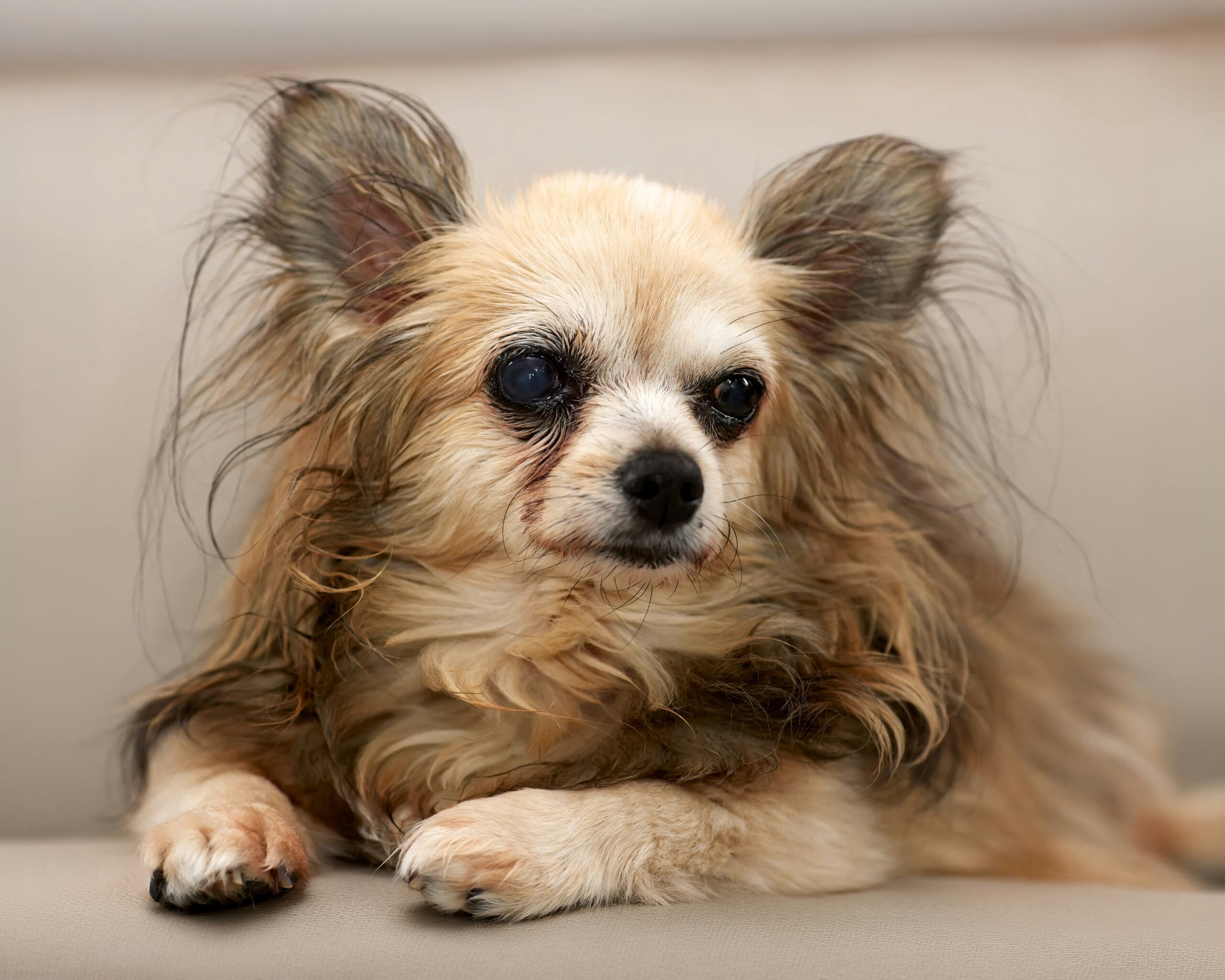
Cataracts in Dogs: Causes, Diagnosis & Treatment
A cloudy-looking eye in an older dog is often assumed to simply be cataracts in dogs, irrespective of cause, but a very common, harmless age-related change looks remarkably similar and is frequently confused with them. This guide explains how to tell the two apart, the real causes of true cataracts including a surprisingly fast-progressing diabetic form, how they’re diagnosed and treated, and what living with a dog experiencing vision loss actually looks like.
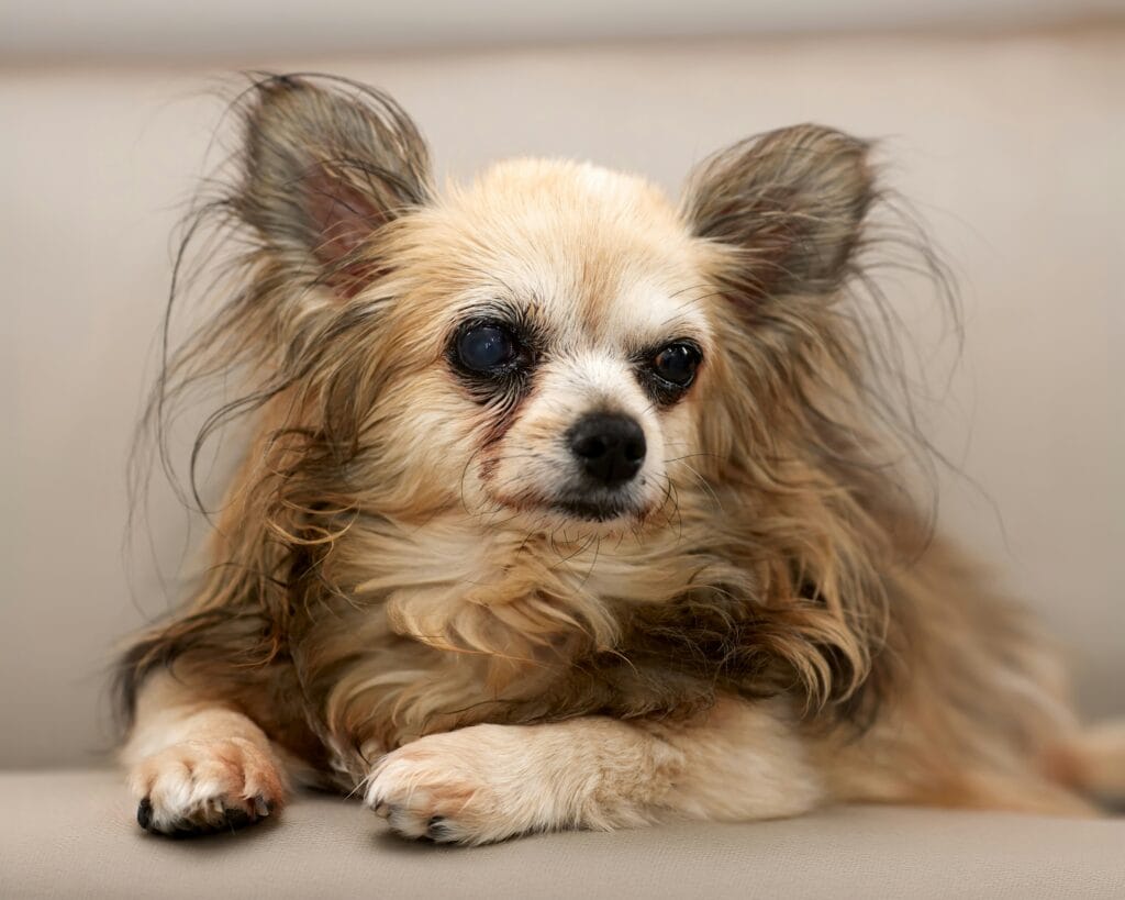
Cataracts in dogs: Cataract vs. Nuclear Sclerosis: A Crucial Distinction
This is genuinely the most important distinction in this entire topic, and the one most frequently confused by owners. Nuclear sclerosis is a normal aging change in the lens, appearing as a subtle bluish-gray haze in both eyes, usually starting around six to eight years of age, and it does not significantly impair vision or require treatment. A true cataract, by contrast, is an actual clouding or opacity within the lens itself, can appear white, and genuinely blocks light from properly reaching the retina, impairing vision to varying degrees depending on how much of the lens is affected. The two can look superficially similar to an untrained eye, which is exactly why a veterinary ophthalmic exam, not a guess based on appearance alone, is needed to tell them apart. Getting this distinction right matters practically: nuclear sclerosis needs no treatment, while a true cataract needs monitoring and, if vision loss is significant, a genuine treatment discussion.
What Causes True Cataracts
Genetics and breed predisposition. Many breeds, including Poodles, Cocker Spaniels, and Boston Terriers, have a notably higher inherited risk of developing cataracts in dogs, sometimes at a relatively young age.
Diabetes mellitus. One of the most significant and fast-acting causes; cataracts in dogs with diabetes commonly develop due to how excess blood sugar affects the lens, sometimes progressing from clear to fully opaque within weeks.
Age-related changes. True cataracts, distinct from harmless nuclear sclerosis, can also develop as part of genuine aging changes in some older dogs.
Trauma or injury. A blow to the eye or penetrating injury can trigger cataract formation in the affected eye.
Congenital cataracts. Some puppies are born with cataracts present from birth, sometimes linked to inherited conditions or, less commonly, an in-utero infection or nutritional issue during pregnancy.
Other eye disease. Chronic inflammation or other underlying eye conditions can sometimes contribute to secondary cataract formation.
| Feature | Nuclear sclerosis | True cataract |
|---|---|---|
| Appearance | Subtle bluish-gray haze | Cloudy, often whiter, more opaque |
| Vision impact | Minimal to none | Ranges from mild to complete vision loss |
| Treatment needed | None | Monitoring; surgery if vision loss is significant |
| Typical onset | Around 6-8+ years, both eyes | Varies widely by cause — can be sudden (diabetes) or gradual |
The Diabetic Cataract Connection
If your dog has diabetes, this section deserves particular attention, since diabetic cataracts represent one of the fastest-progressing forms of the condition. Elevated blood sugar affects the lens through a specific metabolic pathway that draws excess fluid into lens tissue, causing it to swell and cloud, sometimes progressing from a clear lens to complete opacity within just a few weeks of a diabetes diagnosis or a period of poor blood sugar control. This rapid timeline surprises many owners who assume cataracts are always a slow, gradual process. Because of this, veterinarians often recommend regular eye checks specifically for newly diagnosed diabetic dogs, watching closely for the earliest signs of lens changes, since catching and addressing this early gives the best chance at preserving vision through timely surgical intervention if that route is chosen.
Recognizing the Signs
Visible cloudiness in the eye. A whitish or bluish-white appearance to the pupil area, sometimes in one eye first, sometimes both.
Bumping into objects. Particularly noticeable in dim lighting or unfamiliar environments as vision becomes impaired.
Reluctance to navigate stairs or jump. Reduced depth perception affects a dog’s confidence with these movements.
Increased clinginess or hesitation in new spaces. Dogs with declining vision often become more reliant on familiar routes and routines.
Squinting or eye redness. Can occur if the cataract is accompanied by inflammation, which sometimes happens as cataracts mature.
Diagnosis and Treatment
Diagnosis involves a thorough eye exam, often including specialized equipment to examine the lens in detail and distinguish true cataracts from nuclear sclerosis or other eye conditions. There is no effective eye drop or medication that reverses or dissolves an existing cataract, despite various over-the-counter products sometimes marketed for this purpose; surgical removal remains the only treatment that restores vision. Cataract surgery, typically performed by a veterinary ophthalmologist, involves removing the clouded lens and often replacing it with an artificial lens, similar in concept to human cataract surgery, with generally good success rates for otherwise healthy eyes. Not every dog with cataracts is a good surgical candidate, and not every owner chooses surgery, particularly for dogs adapting well without it, making this a genuinely individual decision made together with your veterinarian based on the specific dog’s situation.
Living With a Dog With Vision Loss
Cataract Stages and What They Mean
Veterinary ophthalmologists typically describe cataracts in stages that reflect how much of the lens is affected, and understanding this progression helps make sense of treatment timing discussions. An incipient cataract affects only a small portion of the lens and usually causes minimal noticeable vision impact, often found incidentally during a routine eye exam rather than because an owner noticed symptoms. An immature cataract involves more of the lens and starts to visibly affect vision, though some sight typically remains. A mature cataract affects the entire lens, causing significant to complete vision loss in that eye. A hypermature cataract is a more advanced stage where the lens material has started to break down further, which can sometimes trigger secondary inflammation within the eye requiring closer monitoring or more urgent intervention. Surgery outcomes are often best when performed before a cataract reaches the hypermature stage, which is part of why veterinarians recommend addressing significant cataracts sooner rather than waiting indefinitely once vision loss becomes meaningful.
What Cataract Surgery Actually Involves
Understanding the surgical process itself helps demystify what can otherwise feel like an intimidating decision. The procedure is performed under general anesthesia by a veterinary ophthalmologist, using a technique called phacoemulsification, which uses ultrasound energy to break up and remove the clouded lens material through a small incision, the same core technique used in human cataract surgery. An artificial intraocular lens is often, though not always, implanted to help restore more normal focusing ability afterward. Recovery typically involves an E-collar to prevent rubbing, a course of eye drops administered multiple times daily for several weeks, and follow-up exams to monitor healing and eye pressure. Success rates for appropriately selected candidates are generally good, though as with any surgery, individual factors like overall health and any pre-existing eye conditions affect the specific prognosis discussed with your veterinary ophthalmologist before proceeding.
A Practical Note on Timing
If cataract surgery is a genuine consideration, discussing timing sooner rather than later with your veterinarian generally keeps more options open, since delaying can allow a cataract to progress toward the hypermature stage where outcomes become less predictable. This doesn’t mean every cataract needs immediate surgery, but it does mean an early conversation about the plan is more useful than waiting until vision loss feels urgent.
Frequently Asked Questions
Are all cloudy dog eyes cataracts?
No. Many older dogs have nuclear sclerosis, a normal, harmless haze that doesn’t significantly affect vision and needs no treatment, distinct from true cataracts.
Can diet or supplements prevent or reverse cataracts?
No supplement or diet has been shown to reverse an existing cataract. Some antioxidant supplements are sometimes discussed as potentially supportive for eye health generally, but they are not a treatment for established cataracts.
Is cataract surgery worth it for an older dog?
This depends on the individual dog’s overall health, the severity of vision loss, and owner goals; many older dogs are good surgical candidates and benefit significantly, but it’s a decision made with your veterinarian based on your dog’s specific situation.
How fast do diabetic cataracts develop?
Sometimes remarkably fast, progressing from clear to fully opaque within just a few weeks in some diabetic dogs, which is why regular eye monitoring is recommended after a diabetes diagnosis.
By Dr. Zahid Afzal, DVM — General Veterinary Hospital, Lahore. This article is for general education and does not replace a full physical examination or personalized treatment plan from your own veterinarian.



One Comment