Glaucoma in Dogs: Signs, Emergency Treatment & Prevention
Glaucoma in dogs is one of the true ophthalmic emergencies in veterinary medicine, capable of causing permanent blindness within hours if not treated promptly. This guide explains the difference between primary and secondary glaucoma, why the unaffected eye deserves just as much attention as the painful one, and what emergency and long-term treatment actually involve.
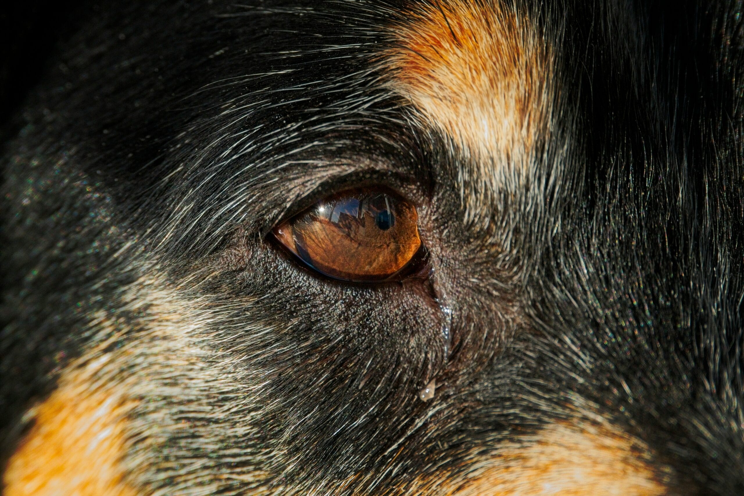
Glaucoma in dogs: primary vs. Secondary Glaucoma
Primary glaucoma in dogs. An inherited structural predisposition affecting how fluid normally drains from the eye, seen more frequently in certain breeds. Because the underlying structural issue is typically present in both eyes, a dog diagnosed with primary glaucoma in one eye faces meaningful risk of eventually developing it in the other eye too, sometimes months to years later.
Secondary glaucoma. Develops as a consequence of another eye condition disrupting normal fluid drainage, including lens luxation (the lens shifting out of position), uveitis (eye inflammation), tumors, or advanced cataracts. Treating the underlying cause is an important part of managing secondary glaucoma, alongside directly addressing the elevated pressure itself.
Why This Is a Genuine Emergency
Elevated pressure inside the eye directly damages the optic nerve and retina, and this damage can become permanent remarkably quickly, sometimes within a matter of hours from significantly elevated pressure. This is fundamentally different from many other eye conditions where a day or two’s delay, while not ideal, doesn’t necessarily change the outcome dramatically. With glaucoma in dogs, that same delay can mean the difference between a treatable eye and permanent, irreversible blindness. This urgency is exactly why any combination of a red, painful, or cloudy-looking eye deserves same-day emergency evaluation, not a wait-and-see approach or a routine appointment scheduled for later in the week.
| Sign | What it suggests |
|---|---|
| Red, bloodshot eye | Inflammation and pressure changes within the eye |
| Cloudy or bluish-hazed cornea | Corneal swelling from elevated pressure |
| Squinting, pawing at the eye | Significant pain from elevated pressure |
| Visibly enlarged eye (advanced/chronic) | Prolonged elevated pressure stretching the eye |
| Sudden vision loss | Optic nerve damage already occurring |
Recognizing the Signs
Redness of the eye. Often one of the more visible early signs, sometimes mistaken for simple conjunctivitis.
Cloudy or blue-hazed appearance. Corneal swelling from the elevated pressure creates a distinctive cloudy look.
Squinting or holding the eye partially closed. A clear pain response.
Rubbing or pawing at the face. A dog attempting to relieve genuine discomfort.
Dilated pupil that doesn’t respond normally to light. A specific, meaningful clinical sign.
Lethargy or reduced appetite. Some dogs show general signs of pain or illness alongside the more obvious eye symptoms.
Diagnosis and Emergency Treatment
What to Do While Traveling to the Emergency Vet
If you suspect glaucoma and are on your way to an emergency clinic, calling ahead so the team can prepare genuinely saves valuable time once you arrive. Keep your dog as calm as possible during transport, since excitement and stress can potentially worsen eye pressure further. Avoid touching or applying any pressure near the affected eye, and don’t attempt any home remedies or over-the-counter eye products, since the wrong product or delay from trying something at home first can cost precious time during a condition where minutes genuinely matter.
Living With a Dog After Vision Loss
If glaucoma does result in partial or complete vision loss despite treatment, many dogs adapt remarkably well, relying more heavily on smell, hearing, and memory of familiar spaces. Keeping furniture and layout consistent, using verbal cues more consistently, and being patient during the adjustment period all support a smooth transition. A blind but pain-free eye, once pressure is controlled, is a genuinely good outcome compared to an eye still causing chronic discomfort, and many owners are surprised by how quickly and completely their dog adapts to navigating the world primarily through senses other than sight.
A Closing Thought
Glaucoma is genuinely one of the conditions where knowledge translates directly into saved vision. Owners who recognize a red, cloudy, or suddenly painful-looking eye as a true emergency, rather than something to watch overnight, give their dog the best possible chance of keeping their sight.
Cost Considerations for Glaucoma Treatment
Emergency glaucoma treatment, including tonometry, emergency medication, and hospitalization if needed, represents a genuine upfront cost, and ongoing daily medication adds a ongoing recurring expense for dogs requiring long-term management. Surgical options, when needed, add a further significant cost. While these figures can feel daunting in the moment, it’s worth weighing them against the alternative: untreated glaucoma leads to permanent blindness and ongoing pain, which itself often requires eventual surgical intervention anyway, just later and after vision is already lost. Discussing the full range of realistic options, including cost, honestly with your veterinarian at the time of diagnosis helps you make an informed decision that fits both your dog’s medical needs and your financial circumstances, rather than facing each decision point as a series of unexpected costs.
How Normal Eye Pressure Works
Understanding the basic mechanics behind eye pressure helps make sense of why glaucoma develops and why treatment focuses where it does. Inside a healthy eye, a clear fluid called aqueous humor is continuously produced and drained in a careful balance, maintaining steady, healthy pressure that keeps the eye properly shaped and functioning. This fluid is produced by structures behind the iris and normally drains out through a specific angle structure where the iris meets the cornea. When this drainage angle is structurally abnormal from birth (as in primary glaucoma) or becomes blocked by inflammation, debris, or a displaced lens (as in secondary glaucoma), fluid continues being produced but can no longer drain properly, and pressure inside the eye rises. This is precisely why treatment approaches target either increasing outflow, decreasing production, or both, since correcting this fundamental imbalance is what actually protects the optic nerve from ongoing damage.
Regular Eye Exams for Predisposed Breeds
For breeds with known predisposition to primary glaucoma, incorporating specific eye pressure checks into routine wellness visits, even before any symptoms appear, offers a genuine opportunity for earlier detection. Some veterinary ophthalmologists recommend baseline tonometry starting at a certain age for predisposed breeds, establishing what’s normal for that individual dog and making any future subtle rise easier to detect and interpret. This proactive screening approach mirrors the same logic used for other silent, serious conditions throughout veterinary medicine, catching a problem while it’s still early and highly treatable is consistently better than waiting for dramatic symptoms to force the issue, and for glaucoma specifically, that early window can be the difference between preserving vision and losing it entirely.
Supporting Your Dog Through Recovery
Whatever the treatment path, the days immediately following diagnosis are often stressful for both dog and owner. Administering eye drops consistently, exactly on schedule, matters genuinely for treatment success, and setting phone reminders or linking doses to existing daily routines helps maintain this consistency even during a busy or emotionally difficult period. An E-collar is often necessary temporarily to prevent your dog from rubbing at an uncomfortable eye, and most dogs adjust to wearing one faster than owners expect. Keeping a simple log of eye appearance, behavior, and any medication given helps you and your veterinary team track progress accurately across follow-up visits, rather than relying on memory during what can be an anxious, fast-moving treatment period.
Working With a Veterinary Ophthalmologist
Given how technical and time-sensitive glaucoma management can be, many general practitioners refer confirmed or complex cases to a veterinary ophthalmologist, a specialist with more advanced diagnostic and surgical tools specifically for eye disease. This referral reflects the genuine complexity of the condition rather than any inadequacy in your regular veterinarian’s initial care, and specialist involvement often makes a meaningful difference in long-term outcomes, particularly for primary glaucoma cases requiring ongoing, carefully calibrated management over years.
Frequently Asked Questions
Can glaucoma in dogs be cured?
Primary glaucoma isn’t curable, since the underlying structural predisposition remains, but pressure can often be managed long-term with medication and monitoring. Secondary glaucoma may improve if the underlying cause is successfully treated.
Is glaucoma painful for dogs?
Yes, elevated eye pressure is genuinely painful, which is part of why prompt treatment matters both for vision and for your dog’s comfort.
Will my dog need surgery?
Not always; many cases are managed with medication alone, though surgery, including eye removal in severe, blind, painful cases, is sometimes necessary depending on how the condition responds to initial treatment.
Which breeds are most at risk for primary glaucoma?
Certain breeds show notably higher predisposition due to inherited eye drainage structure differences; your veterinarian can advise on your specific breed’s risk level and whether preventive monitoring is recommended.
By Dr. Zahid Afzal, DVM — General Veterinary Hospital, Lahore. This article is for general education and does not replace a full physical examination or personalized treatment plan from your own veterinarian. If your dog’s eye appears red, cloudy, or painful, seek emergency care immediately.